Health Science Programs receive cutting-edge equipment
The Diagnostic Medical Sonography program and the Radiologic Technology program at Gadsden State Community College enhanced its training capabilities with the addition of cutting-edge equipment. DMS received two ultrasound simulators while Rad Tech added a refurbished x-ray machine to the classroom.
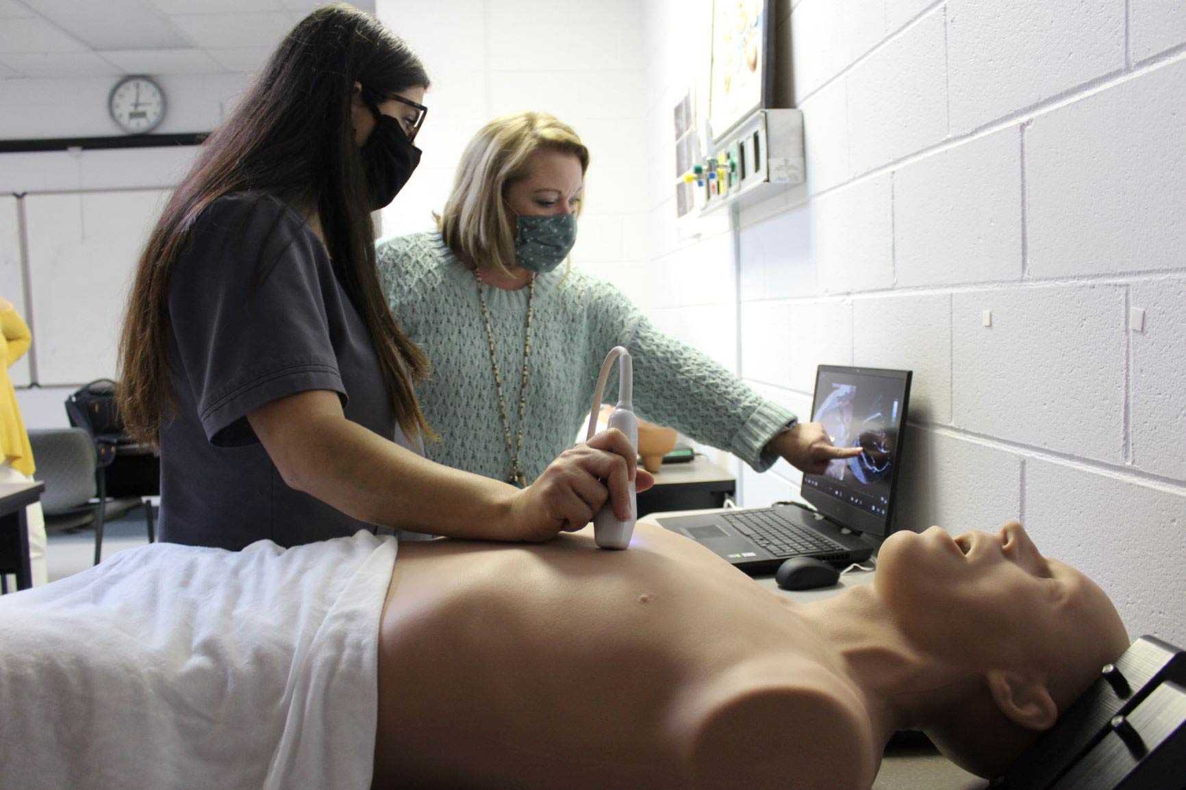
DMS ultrasound simulators
The ultrasound simulators, which are on a manikin-based system, are known as Bob and Catherine and are located in the DMS lab on the Valley Street Campus.
“Our students are used to scanning live patients in our school lab, but few of them have any abnormalities,” said Rebecca Southern, DMS program director. “Bob and Catherine let the students see abnormalities under our guidance so they can identify certain pathologies.”
The high-fidelity ultrasound training simulators facilitate the learning processes for cardiac, lung, abdominal and obstetrics/gynecology all on one platform.
“They assist in teaching the skills for ultrasound probe handling, image interpretation, diagnoses and clinical decision-making,” she said.
“Bob” assists in teaching students both transthoracic and transesophageal echocardiography. He also helps them learn to perform abdominal scans.
“Bob has 109 different pathologies for the students to view and access, including aneurysms, masses and tumors,” Southern said. “He can have gallstones, valve issues, hernias. There are so many opportunities for our students to find, identify and label pathologies.”
“Catherine” teaches the students how to perform obstetric and gynecologic ultrasound exams using an endocavity probe or a transabdominal probe. She has 41 pathologies.
“Through Bob and Catherine, our students can see organs, tissues and vessels as well as potential pathologies,” she said. “This is a great way for our students to learn sectional anatomy. They are learning how these organs are layered in the body and better understand their relationship to one another on the ultrasound image.”
The ultrasound training simulators are produced by CAE Vimedix, a global company that offers integrated education and training solutions to healthcare students. David Wildermuth, a company representative, said Gadsden State is one of only a few community colleges nationwide to use their product.
“While about 20 other community colleges use Vimedix, Gadsden State is the only community college that has the fully-loaded Vimedix simulators with all of our modules that can be used across multiple specialties,” he said.
Southern is excited to have the $140,000 simulators that have allowed her to use a pathology learning platform that is transferrable to the classroom or an online teaching environment if necessary.
“The DMS program at Gadsden State has only been in existence for three years, but we have been able to secure some of the best ultrasound equipment available to colleges and universities,” she said. “And, it’s paying off. We have had three graduating classes, with successful outcomes, in which our graduates are making a positive impact in the sonography industry.”
 |
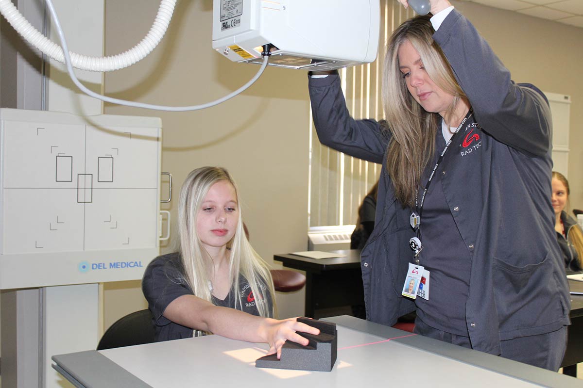
Rad Tech X-ray machine
Gadsden State is now the only community college in the state to have an x-ray machine in the classroom, which is a huge benefit for the students.
“The radiographic room allows the clinical coordinator, Ashley Crusey, and I to easily demonstrate a concept by simply refocusing the students’ attention to the side of the classroom and actually showing the procedure,” said Dr. Joey Battles, program director.
Battles, who has been teaching for 26 years, said having a classroom radiographic room has always been a dream.
“Positioning of a patient for an examination can be difficult to see in a photograph in a book or on a PowerPoint slide,” he said. “If students are having trouble understanding what we are discussing, we can take them to the machine and simply show them.”
Holli Shaver of Summerville, Ga., said her level of understanding has increased since the installation of the x-ray machine.
“When discussing different positions or projections in class, I can better understand what I have learned through visualizing the position of the patient or part while it’s being taught to me,” she said. “I can manipulate the tube how I need to in order to put all the pieces of the puzzle together.”
The radiographic room helps solidify concepts for the students, especially those who are visual and/or hands-on learners.
“I learn more from experience and prefer to be physically engaged in the learning process,” said Corley Groover, a student from Rainbow City. “The machine gives me the opportunity to learn from trial and error so I can understand and fix my mistakes.”
Fellow student Brenna Simpson of Attalla is also a hands-on learner.
“I’m able to retain positions and angulations better,” she said. “In the end, I’m better prepared for my exams, and I’ll be better prepared for the workforce.”
 |